
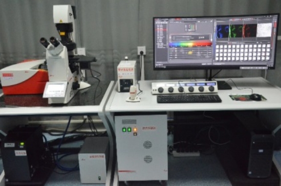
Microscopic Imaging Room
Microscopic Imaging Room (room 308)
The Microscopic Imaging Room(1)provides scientific services, which mainly include the services of preparation of paraffin sections, conventional tableting, dyeing, drying, decolorization, micrography, microdissection, microscopic live imaging, and fluorescence imaging. The equipment in the microscope room consists of the laser scanning confocal microscope, a fluorescence microscope, and paraffin sectioning equipment.
Laser Scanning Confocal Microscope
Basic Information:
Model: TCS SP8 SR
Manufacturer: Leica (Germany)
Device Configuration:
Microscope: DMi8 full-electric inverted microscope
Observation method: bright field, differential interference contrast, polarization, fluorescence (Filter: UV/GFP/RFP)
Fluorescent lighting: long-life metal halide light source, life of more than 2000 hours, fiber optic conduction
Objective: semi apochromatic objective 1.25 x; 10 x, 20 x, 40 x, 63 x, 100 x confocal dedicated objective lens
Laser: 405 near-ultraviolet, 488 blue, 514 green, 552 yellow, 638 red solid-state laser
Detectors: 2 Leica patented spectral high sensitivity detectors, 3 PMT conventional spectral detectors
Spectroscopic scanning function: all 5 fluorescence detectors can be used for spectral scanning, and the range of spectral scanning is 400-800 nm
The maximum scanning resolution is 8192x8192 pixels. gray scale: 8, 12, and 16 bits
It can carry out multi-dimensional combined scanning, including X, Y, Z, T, λ (spectral wavelength), θ (rotation angle), I (light intensity), A (region), etc., realizing point, line, curve, area, and spectral wavelength scanning and so on. Five fluorescent signals plus one transmitted light can be collected simultaneously
Test Project:
Observation of all kinds of stained, non-stained, fluorescence-labeled tissue sections and living cells. Accurate description, localization as well as qualitative, quantitative, and periodic analysis of dynamic changes of the above structures
Qualitative, quantitative, periodic, and localized observation of cellular biological substances
Direct observation and dynamic characterization of cell ion channels
3D image reconstruction
Co-localization analysis
Analysis of dynamic fluorescence intensity, ratio value measurement (calcium ion)
Fluorescence recovery after photobleaching, fluorescence resonance energy transfer
Automatic Upright Fluorescence Microscope
Basic Information:
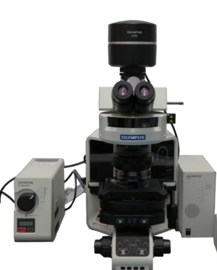
Model: BX63
Manufacturer: Olympus (Japan) Company
Device Configuration:
Observation method: Bright field\Phase\Fluorescence\DIC
Objective: plan semi apochromat objective 4×, 10×, 20×, 40×, 40× (Phase), 60× (Oil), 100× (Oil)
Semiconductor monochromatic cold CCD:2/3 inches, physical pixels 1.4 million; Semiconductor color cold CCD:2/3 inches,12.5 million pixels, image acquisition speed: 15 p/ SEC
Filter: UV/ GFP/ RFP/YFP\
Test Project:
1. Observation of general biological staining slice
2. Research of plants' living molecular markers
3. Research of cell immunofluorescence
Upright Fluorescence Microscope
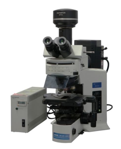
Upright Fluorescence Microscope
Basic Information:
Model: BX51
Manufacturer: Olympus (Japan) Company
Device Configuration:
1. Observation method: Bright field\Fluorescence
2. Objective: plan semi apochromat objective 4×, 10×, 20×, 40×, 100×
3. CCD: cold color CCD: 2/3 inch, 12.5 million pixels, image acquisition speed: 15 p/ SEC
4. Filter: UV/ GFP/RFP
Test Project:
1. Observation of general biological staining slice
2. Research of plants' living molecular markers
3. Research of cell immunofluorescence
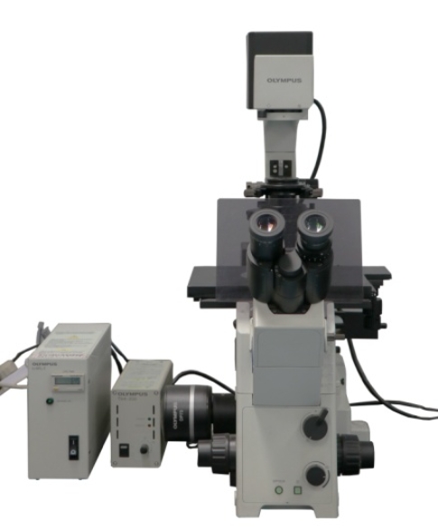
Inverted fluorescence Microscope
Basic Information:
Model: IX71
Manufacturer: Olympus (Japan) Company
Device Configuration:
1. Observation method: Bright field \ Phase\Fluorescence
2. Objective:long working distance fluorescence objective 4×, 10×, 20×, 40×
3. CCD: cold color CCD: 17 million pixels, image acquisition speed: 15 p/SEC
4. Filter: GFP/RFP
Test Project:
1. Observation of biocytoculture
2. Research into biological tissue culture
3. Observation of living cell fluorescence
Inverted fluorescence Microscope
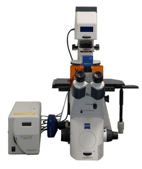
Basic Information:
Model: Axio Observer D1
Manufacturer: Carle Zeiss(Germany) Company
Device Configuration:
1. Observation:Bright field \ Phase\Fluorescence\DIC\Polarized light.
2. Eyepiece: 10X
3 .Objective: Objective LD A-Plan 5×|0.15 Ph1
Objective EC Plan-Neofluar 10×|0.3 Ph1
Objective LD A-Plan 20×|0.35 Ph1
Objective Plan-Apochromat 20×|0.8
Objective LD Plan-Neofluar 40×|0.6 Corr Ph2
Objective EC Plan-Neofluar 40×|1.30 Oil
Objective Plan-Apochromat 63×|1.40 Oil
Objective Plan-Apochromat 100×|1.4 Oil
4.CCD: color digital CCD 5 million effective physical pixels
Cold monochrome CCD Two million eight hundred and thirty thousand effective physical pixels.
5.Filter: UV EX BP 365/12, BS FT 395, EM LP 397
RFP EX BP 450-490, BS FT 510, EM LP 515
GFP EX BP 510-560, BS FT 580, EM LP 590
YFP EX BP 500|20, BS FT 515, EM BP 535|30
Test Project:
1. Observation of biocytoculture
2. Research into biological tissue culture
3. Observation of living cell fluorescence
Stereoscopic Fluorescence Microscope
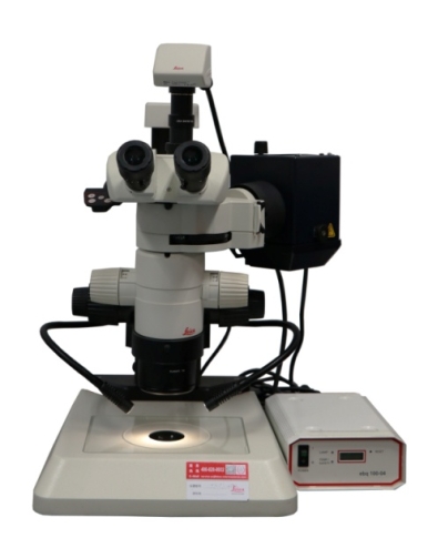
Basic Information:
Model: LEICA MZ10F
Manufacturer: LEICA (Germany) Company
Device Configuration:
1.Observation method: Stereo FluorescenceMagnification range: 8x-80x
2.Eyepiece: 10×/23B
3.Objective: apochromat objective1.0×
4.CCD: Leica DFC450 C Digital Camera
5.Filter: UV/GFP/RFP
Test Project:
1. Plant organ dissection and observation
2. Biological section observation
3. Biological specificity of a fluorescent protein
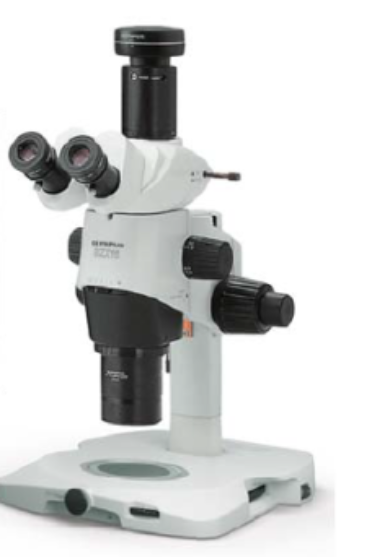 Continuous Vaploid Fluorescence Micrograph System
Basic Information:
Model: SZX16
Manufacturer: Olympus (Japan)
Device Configuration:
1. Observation method: Incident and reflected light from a bright field
2. Objective: High-resolution APO objective 1×
3. CCD: Color CCD, 18 million pixels, image acquisition speed: 15 p/SEC
Test Project:
1. Plant organ dissection and observation
2. Biological section observation
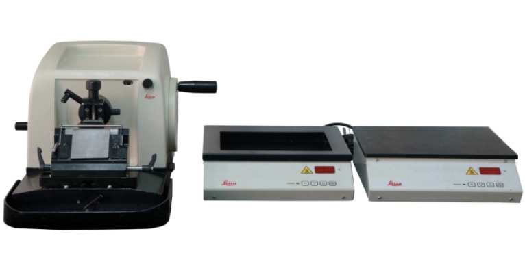
Rotary Microtome
Basic Information:
Model: RM2016
Manufacturer: LEICA (Germany) Company
Device Configuration:
1.Slice thickness from 1-60um
2.Samples of horizontal displacement of 25mm
3.Samples vertical maximum 70mm in diameter
Test Project:
Horticultural crops' paraffin section
Organization stand
Baking organization
Semi-Thin Microtome
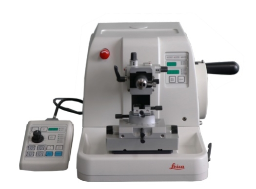
Basic Information:
Model: RM2265
Manufacturer: LEICA (Germany) Company
Device Configuration:
1. Slice thickness 0.25-100μm
2. Vertical distance 70mm
3. Electric injection speed ≥2
Test Project:
Semi-thin as observed by electron microscopy and staining of plant tissue structure.